GDA Nursing Class Note 11
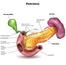
- Pancreas:
- Location: The pancreas is an organ located behind the stomach, in the upper abdomen.
- Structure: It is a glandular organ with both endocrine and exocrine functions.
- Endocrine Function: The pancreas produces important hormones like insulin and glucagon, which play a crucial role in regulating blood sugar levels.
- Exocrine Function: The pancreas produces pancreatic juices containing digestive enzymes that are released into the small intestine to aid in the digestion of carbohydrates, proteins, and fats.
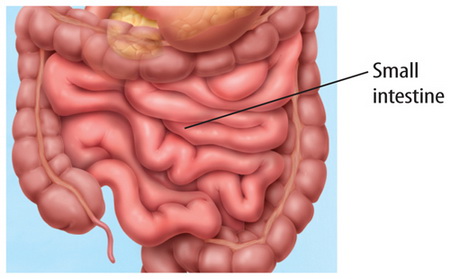
- Small Intestine:
- Location: The small intestine is the longest part of the digestive tract and is located between the stomach and the large intestine.
- Structure: It is divided into three segments: duodenum, jejunum, and ileum.
- Digestion and Absorption: The small intestine is the primary site for the digestion and absorption of nutrients from food. Enzymes from the pancreas and bile from the liver aid in this process.
- Villi and Microvilli: The inner lining of the small intestine is covered with tiny finger-like projections called villi, which increase the surface area for nutrient absorption. Each villus is further covered with even smaller projections called microvilli.
- Nutrient Transport: Nutrients, such as sugars, amino acids, and fatty acids, are absorbed through the walls of the small intestine and then transported to the bloodstream to be distributed throughout the body.
- Large Intestine (Colon):
- Location: The large intestine is the final part of the digestive system and is located in the lower abdomen.
- Structure: It is wider in diameter but shorter than the small intestine.
- Functions: The main functions of the large intestine are to absorb water and electrolytes from indigestible food residues, store and concentrate fecal matter, and house a diverse population of beneficial gut bacteria.
- Ascending Colon:
- Location: The ascending colon is the first segment of the large intestine and is located on the right side of the abdomen.
- Path: It begins at the cecum, where it connects to the small intestine, and extends upward towards the liver and then makes a turn called the right colic or hepatic flexure.
- Function: In the ascending colon, water and electrolytes are absorbed from the remaining indigestible food particles, and the material becomes more solid as it progresses through the colon.
- Transverse Colon:
- Location: The transverse colon is the second segment of the large intestine and is situated horizontally across the upper abdomen.
- Path: It starts at the right colic flexure (where the ascending colon turns) and continues to the left colic or splenic flexure.
- Function: In the transverse colon, the process of absorbing water and electrolytes continues, and some remaining nutrients may also be absorbed.
- Descending Colon:
- Location: The descending colon is the third segment of the large intestine and is located on the left side of the abdomen.
- Path: It begins at the left colic flexure and travels downward.
- Function: In the descending colon, any remaining water and electrolytes are absorbed, and the stool becomes more solid as it approaches the last segment, the sigmoid colon.
- Sigmoid Colon:
- Location: The sigmoid colon is the final S-shaped segment of the large intestine, located in the lower left abdomen.
- Path: It starts at the end of the descending colon and connects to the rectum.
- Function: In the sigmoid colon, the stool is further dehydrated, and it is stored until it is ready to be eliminated from the body during a bowel movement.
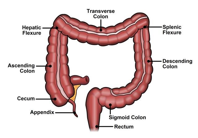
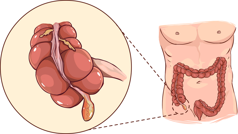
Appendix:
- Location: The appendix is a small, finger-shaped pouch attached to the cecum, which is the first part of the large intestine. It is situated in the lower right abdomen.
- Structure: The appendix does not have any significant digestive function and is often considered a vestigial organ. It is about 2 to 4 inches (5 to 10 cm) long.
- Uncertain Function: While the exact function of the appendix in the human body is not entirely clear, it is believed to have some roles related to the immune system and gut health. It may serve as a reservoir for beneficial gut bacteria, helping to repopulate the intestines after a bout of diarrhea or other gastrointestinal disturbances.
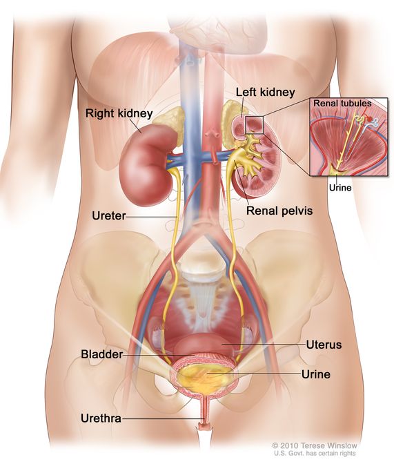
- Kidneys:
- Location: The kidneys are two bean-shaped organs located on either side of the spine in the upper part of the abdomen, just below the ribcage.
- Function: The kidneys play a vital role in maintaining the body’s internal environment. Their primary functions include:
- Filtration: The kidneys filter waste products, excess salts, and toxins from the blood, forming urine.
- Regulation of Water and Electrolyte Balance: The kidneys control the body’s water and electrolyte levels by selectively reabsorbing essential substances back into the bloodstream while eliminating excess through urine.
- Acid-Base Balance: The kidneys help regulate the body’s pH by excreting or reabsorbing hydrogen ions and bicarbonate ions.
- Blood Pressure Regulation: Through the renin-angiotensin-aldosterone system, the kidneys play a role in regulating blood pressure.
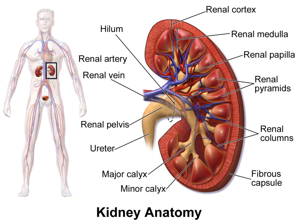
- Urinary System: The kidneys are part of the urinary system, which also includes the ureters (tubes that transport urine from the kidneys to the bladder), the urinary bladder (where urine is stored), and the urethra (through which urine is eliminated from the body).
- Capsule:
- General Definition: A capsule is a protective and often fibrous covering that surrounds certain organs or structures, providing support and maintaining their shape.
- Cortex:
- General Definition: The cortex refers to the outer layer of an organ or structure, usually having a distinct appearance and function from the inner layers.
- Medulla:
- General Definition: The medulla refers to the innermost part of an organ or structure, often having different functions from the outer layers.
- Pelvis:
- General Definition: The pelvis is a bony structure that forms the base of the spine and connects the spine to the lower limbs. It also houses and protects some of the body’s reproductive and digestive organs.
Blood circulation in kidneys:
- Renal Arteries: The kidneys receive blood supply through the renal arteries, which branch off from the abdominal aorta, the main artery of the body. There is one renal artery for each kidney.
- Afferent Arterioles: The renal arteries divide into smaller arterioles within the kidneys, and each arteriole leads to a structure called the glomerulus.
- Glomerulus: The glomerulus is a tuft of capillaries surrounded by a structure called Bowman’s capsule. Blood is filtered through the glomerulus into the Bowman’s capsule, where the process of urine formation begins.
- Filtration: As blood passes through the glomerulus, small molecules like water, electrolytes, waste products, and some nutrients are filtered out from the blood into the Bowman’s capsule. This initial filtrate is known as the “glomerular filtrate.”
- Regulation of Blood Flow: The blood flow in the kidneys is tightly regulated to ensure proper filtration and maintenance of fluid and electrolyte balance. Hormones such as renin and aldosterone, as well as the autoregulation of blood flow through the kidney’s juxtaglomerular apparatus, play a crucial role in regulating kidney function and blood pressure.
- Waste Removal: Along with reabsorption, the peritubular capillaries and vasa recta also help in removing waste products that were not reabsorbed during filtration. These waste products are carried away from the kidneys to be eventually excreted as urine.
Lorem ipsum dolor sit amet, consectetur adipiscing elit. Ut elit tellus, luctus nec ullamcorper mattis, pulvinar dapibus leo.
Lorem ipsum dolor sit amet, consectetur adipiscing elit. Ut elit tellus, luctus nec ullamcorper mattis, pulvinar dapibus leo.