GDA Nursing Class note 9
Sense organs are specialized structures in living organisms that detect and respond to various stimuli from the environment. They play a crucial role in gathering information about the surroundings, enabling organisms to interact with and adapt to their surroundings. Here are some key points about the sense organs:
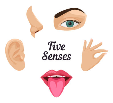
- Vision (Eyes): The eyes are the sense organs responsible for the sense of sight. They detect light and convert it into electrical signals that are transmitted to the brain for interpretation.
- Hearing (Ears): The ears are the sense organs responsible for the sense of hearing. They detect sound waves and convert them into electrical signals that are interpreted by the brain.
- Smell (Nose): The nose is the sense organ responsible for the sense of smell. It contains olfactory receptors that detect and distinguish different odors in the environment.
- Taste (Tongue): The tongue is the sense organ responsible for the sense of taste. It contains taste buds that can detect five primary tastes: sweet, sour, salty, bitter, and umami (savory).
- Touch (Skin): The skin is the largest sense organ in the human body and is responsible for the sense of touch. It contains various receptors that detect pressure, temperature, pain, and other tactile sensations.
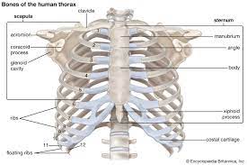
Ribs:
Ribs are a set of long, curved bones that form the rib cage, which protects the vital organs in the chest, such as the heart and lungs. Here are some key points about ribs:

- Structure: The human rib cage consists of 12 pairs of ribs, collectively known as the thoracic cage. Each rib is a thin, flat bone that curves around the chest, with the shape resembling a shallow arc. The ribs are connected to the thoracic vertebrae in the back and either directly or indirectly attach to the sternum (breastbone) in the front.
- Types of Ribs: The 12 pairs of ribs can be classified into three types based on their attachment to the sternum:
- True Ribs: The first seven pairs of ribs (ribs 1-7) are true ribs. They are directly connected to the sternum via costal cartilage, which is a flexible connective tissue.
- False Ribs: The next three pairs of ribs (ribs 8-10) are false ribs. They do not directly connect to the sternum but instead attach to the cartilage of the rib above.
- Floating Ribs: The last two pairs of ribs (ribs 11-12) are floating ribs. They are not connected to the sternum or the rib above and are only attached to the vertebrae in the back.
- Functions:
- Protection: Ribs provide a protective cage around the thoracic organs, including the heart, lungs, and major blood vessels. They help shield these vital organs from injury.
- Support: Ribs contribute to the structural support of the chest, assisting in maintaining the shape and stability of the thoracic cavity.
- Breathing: The movement of ribs, along with the muscles between them, plays a crucial role in the process of breathing. The ribs expand and contract during inhalation and exhalation, facilitating the expansion and contraction of the lungs.
- Rib Injuries:
- Fractures: Rib fractures can occur due to trauma, such as from a fall, impact, or strong compression. Fractured ribs can be painful and may interfere with breathing and movement.
- Dislocation: Rib dislocation is rare but can happen if a rib slips out of its normal position, typically at the joint where it attaches to the thoracic vertebrae.
- Costochondritis: Costochondritis is the inflammation of the cartilage that connects the ribs to the sternum. It can cause chest pain, particularly during movements and deep breathing.
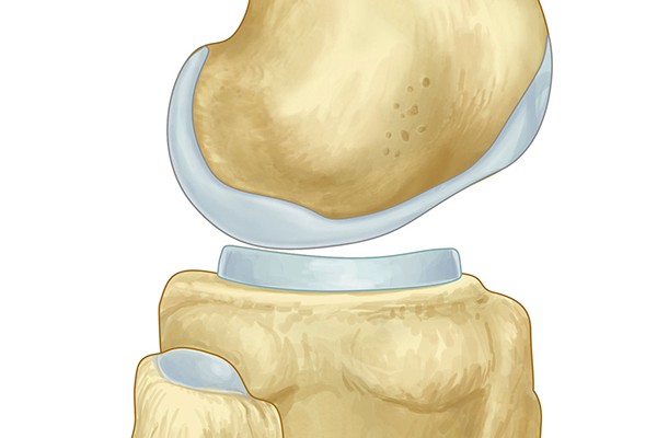
Cartilage is a flexible connective tissue found in various parts of the body. It provides support, cushioning, and smooth surfaces for joint movement. Here are some key points about cartilage:
- Structure: Cartilage is composed of cells called chondrocytes embedded in a gel-like matrix. The matrix consists of collagen fibers, proteoglycans (proteins with attached sugar molecules), and water. The composition of the matrix gives cartilage its unique properties.
The vertebral column, also known as the spinal column or backbone, is a flexible and intricate structure that forms the central axis of the skeleton in vertebrates, including humans. Here are some key points about the vertebral column:
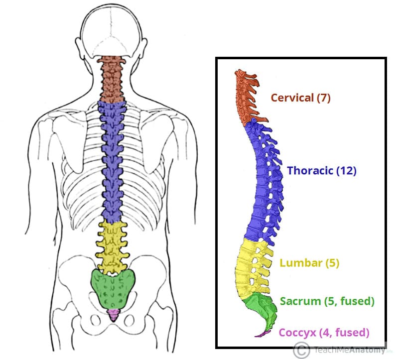
- Structure: The vertebral column is composed of a series of individual bones called vertebrae. In humans, there are typically 33 vertebrae, which are stacked on top of each other in a column-like arrangement.
- Regions of the Vertebral Column: The vertebral column can be divided into five regions, each with a specific number of vertebrae:
- Cervical Vertebrae (C1-C7): Located in the neck region, these vertebrae support the head and allow for a wide range of head movements.
- Thoracic Vertebrae (T1-T12): These vertebrae are associated with the rib cage and provide attachment points for the ribs.
- Lumbar Vertebrae (L1-L5): Found in the lower back, the lumbar vertebrae are the largest and strongest vertebrae, providing support for the upper body and facilitating movements like bending and twisting.
- Sacral Vertebrae (S1-S5): Fused together, the sacral vertebrae form the sacrum, a triangular bone at the base of the vertebral column. The sacrum connects the vertebral column to the pelvic bones.
- Coccygeal Vertebrae (Co1-Co4): Also known as the coccyx or tailbone, these small, fused vertebrae are located at the end of the vertebral column.
- Intervertebral Discs: In between each vertebra, there are intervertebral discs made of cartilage. These discs act as shock absorbers and provide flexibility and cushioning between the vertebrae.
- Functions:
- Support and Stability: The vertebral column provides structural support and stability to the body, maintaining an upright posture and protecting the spinal cord.
- Protection of the Spinal Cord: The vertebral column surrounds and protects the spinal cord, which is a vital component of the central nervous system.
- Movement: The flexibility of the vertebral column allows for a wide range of movements, including bending, twisting, and extension of the trunk.
- Attachment Sites: The vertebrae serve as attachment points for muscles, ligaments, and tendons, allowing for movement and stability of the spine.
- Curvatures of the Vertebral Column: The vertebral column has natural curvatures that contribute to its strength and flexibility. There are four main curvatures:
- Cervical Curve: A slight inward curve in the neck region.
- Thoracic Curve: An outward curve in the upper back region.
- Lumbar Curve: An inward curve in the lower back region.
- Sacral Curve: An outward curve in the sacral region.
- Spinal Disorders and Conditions:
- Scoliosis: Abnormal lateral curvature of the spine, which can cause an uneven appearance of the back.
- Herniated Disc: A condition where the intervertebral disc protrudes and puts pressure on nearby nerves, resulting in pain and other symptoms.
- Spinal Stenosis: Narrowing of the spinal canal, leading to compression of the spinal cord and nerves.
- Degenerative Disc Disease: Age-related wear and tear of the intervertebral discs, causing pain and decreased mobility.
- Understanding the structure and function of the vertebral column is crucial for healthcare professionals, including orthopedic surgeons, physical therapists, and chiropractors, to diagnose and treat spinal conditions and ensure optimal spine health.