GDA Nursing Class Note 10
Lungs are a pair of vital organs located in the chest cavity of humans and many other animals. They play a crucial role in the respiratory system by facilitating the exchange of oxygen and carbon dioxide between the body and the environment. Here are some key points about the lungs:
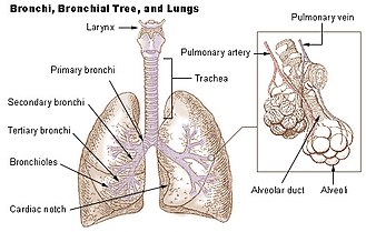
- Anatomy: The lungs are spongy, cone-shaped organs situated on either side of the heart within the rib cage. They are divided into lobes, with the right lung consisting of three lobes (upper, middle, and lower) and the left lung having two lobes (upper and lower). The lungs are enclosed by a protective membrane called the pleura.
- Function: The primary function of the lungs is respiration. During inhalation, air enters the body through the nose or mouth, travels down the windpipe (trachea), and enters the lungs through the bronchial tubes. Inside the lungs, the bronchial tubes branch out into smaller passageways called bronchioles, which ultimately lead to tiny air sacs called alveoli. It is in the alveoli where the exchange of oxygen and carbon dioxide takes place.
- Gas Exchange: The alveoli are surrounded by an extensive network of capillaries. Oxygen from the inhaled air diffuses across the thin walls of the alveoli into the capillaries, where it binds with red blood cells. At the same time, carbon dioxide, a waste product of cellular metabolism, moves from the capillaries into the alveoli and is then expelled during exhalation.
- Breathing Process: The lungs are involved in the process of breathing, which consists of inhalation and exhalation. When you breathe in, the diaphragm (a dome-shaped muscle below the lungs) contracts and moves downward, while the muscles between the ribs also expand. This action increases the volume of the chest cavity, causing air to rush into the lungs. During exhalation, the diaphragm and intercostal muscles relax, reducing the chest cavity’s size and forcing air out of the lungs.
The heart is a vital organ that serves as the main pump of the circulatory system, supplying oxygen and nutrients to the body’s tissues. Here is a comprehensive explanation of the heart:
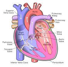
- Anatomy: The heart is a muscular organ located in the chest cavity, slightly to the left of the midline. It is about the size of a clenched fist and is enclosed in a protective sac called the pericardium. The heart has four chambers: two atria (singular: atrium) and two ventricles. The atria receive blood returning to the heart, while the ventricles pump blood out of the heart.
- Structure and Function of Chambers: The right atrium receives deoxygenated blood from the body through two large veins called the superior vena cava (from the upper body) and inferior vena cava (from the lower body). From the right atrium, blood flows into the right ventricle, which then pumps the deoxygenated blood to the lungs for oxygenation through the pulmonary artery. Oxygenated blood returns to the heart from the lungs via the pulmonary veins, entering the left atrium. From there, blood moves into the left ventricle, which forcefully pumps oxygenated blood to the rest of the body through the aorta.
- Valves: The heart has four valves that ensure one-way blood flow through the chambers. The tricuspid valve is located between the right atrium and right ventricle, while the mitral (bicuspid) valve is situated between the left atrium and left ventricle. The pulmonary valve guards the entrance to the pulmonary artery, and the aortic valve is positioned at the beginning of the aorta. These valves open and close synchronously, preventing the backward flow of blood.
- Coronary Circulation: The heart itself requires a continuous supply of oxygen and nutrients. Coronary arteries, arising from the base of the aorta, encircle the heart and provide it with oxygenated blood. The coronary veins collect deoxygenated blood and drain it into the right atrium.
- Electrical Conduction: The heart has its internal electrical system that coordinates its contractions. The sinoatrial (SA) node, located in the right atrium, initiates the electrical impulses, causing the atria to contract. The impulses then travel to the atrioventricular (AV) node, located between the atria and ventricles, which relays the signals to the ventricles, causing them to contract and pump blood.
- Cardiac Cycle: The cardiac cycle refers to the complete sequence of events that occur in the heart during one heartbeat. It consists of two phases: diastole and systole. During diastole, the heart muscles relax, the atria fill with blood, and the ventricles receive blood from the atria. In systole, the atria contract to push the remaining blood into the ventricles, and then the ventricles contract, pumping blood out of the heart.
- Circulatory System: The heart is part of the circulatory system, which includes blood vessels (arteries, veins, and capillaries). Arteries carry oxygenated blood away from the heart to the body’s tissues, while veins transport deoxygenated blood back to the heart. Capillaries are tiny, thin-walled vessels where the exchange of oxygen, nutrients, and waste products occurs between the blood and the surrounding tissues.
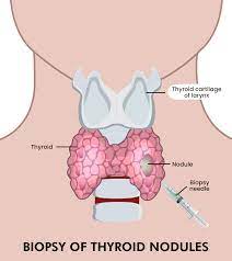
The thyroid gland is a small, butterfly-shaped gland located in the front of the neck, just below the Adam’s apple. It plays a crucial role in regulating various bodily functions by producing and releasing hormones. Here are some key points about the thyroid gland:
- Anatomy: The thyroid gland consists of two lobes connected by a narrow bridge of tissue called the isthmus. It is located in the lower part of the neck, wrapped around the trachea (windpipe). The thyroid is composed of numerous follicles that contain thyroid cells responsible for hormone production.
- Hormone Production: The thyroid gland produces and releases two main hormones: thyroxine (T4) and triiodothyronine (T3). These hormones contain iodine and are essential for regulating metabolism, growth, development, and body temperature. The production of T4 and T3 is regulated by the pituitary gland, which releases thyroid-stimulating hormone (TSH) to stimulate the thyroid.
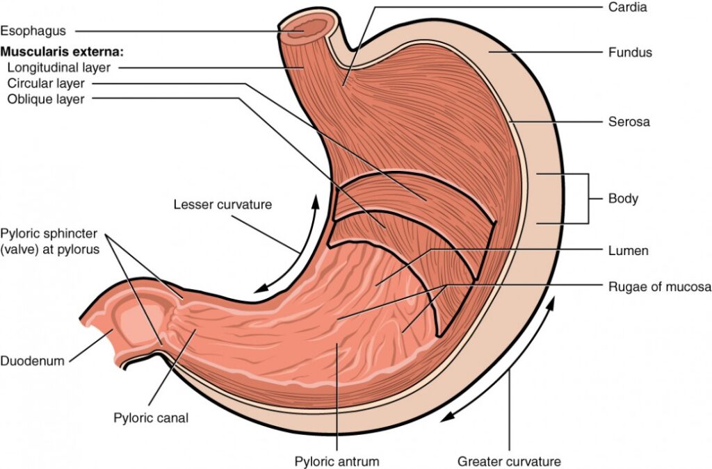
Stomach:–
The stomach is an important organ in the digestive system responsible for breaking down food and aiding in the absorption of nutrients. Here are some key points about the stomach:
- Anatomy: The stomach is a muscular, J-shaped organ located in the upper abdomen, just below the rib cage. It connects to the esophagus (food pipe) at the upper end and the small intestine at the lower end. The stomach has four main regions: the cardia (where the esophagus connects), the fundus (upper portion), the body (central region), and the pylorus (lower portion).
- Function: The stomach has several functions in the digestive process. It receives food from the esophagus and mixes it with digestive juices, breaking it down into smaller particles called chyme. The stomach secretes gastric juices, including hydrochloric acid and enzymes like pepsin, which help in the digestion of proteins. It also serves as a reservoir, storing food until it is ready to be further digested and absorbed in the small intestine.
- Digestive Process: When food enters the stomach, the muscles of the stomach walls contract and relax, churning and mixing the food with gastric juices. This process, known as mechanical digestion, helps break down the food into smaller particles and mixes it with digestive enzymes. The acidic environment of the stomach aids in the chemical digestion of proteins. The partially digested food, now called chyme, gradually moves into the small intestine for further digestion and absorption.
- Hormonal Regulation: The stomach plays a role in the hormonal regulation of digestion. The presence of food in the stomach stimulates the release of the hormone gastrin, which triggers the secretion of gastric juices. Gastrin also promotes stomach contractions, aiding in the mixing and movement of food.
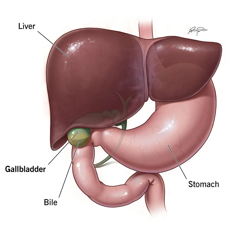
The gallbladder is a small, pear-shaped organ located beneath the liver. It plays a crucial role in the digestive system by storing and concentrating bile produced by the liver. Here are some key points about the gallbladder:
- Anatomy: The gallbladder is located on the right side of the abdomen, tucked beneath the liver. It is connected to the liver and the small intestine by a series of ducts called the bile ducts. The gallbladder has a capacity of around 30 to 50 milliliters and consists of three main parts: the fundus, body, and neck.
- Function: The primary function of the gallbladder is to store and concentrate bile, a yellowish-green fluid produced by the liver. Bile is essential for the digestion and absorption of fats in the small intestine. When food, especially fatty foods, enters the small intestine, the gallbladder contracts and releases stored bile into the digestive tract through the bile ducts.